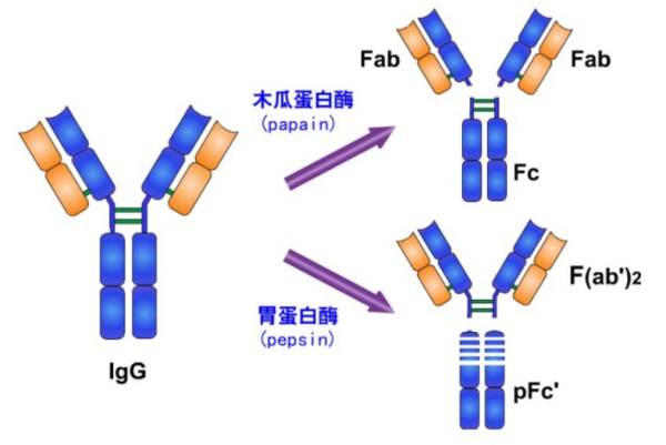
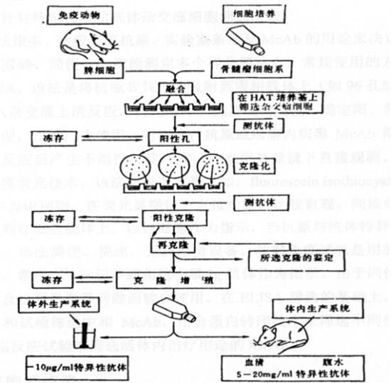
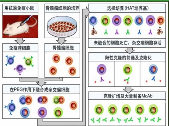
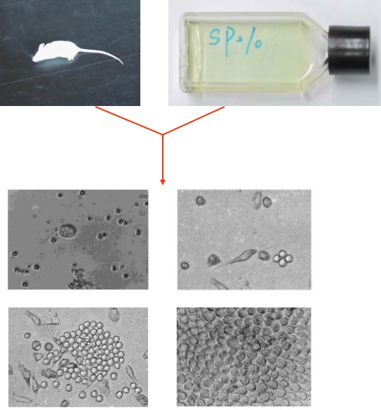
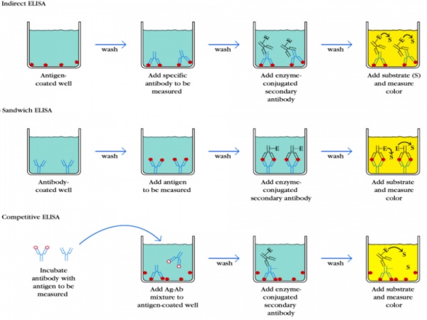
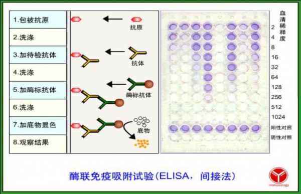
Antibody concept After the animal body is stimulated by the antigenic substance, the B lymphocyte is transformed into an immunoglobulin produced by the plasma cell and capable of specifically binding to the corresponding antigen. Such an immunoglobulin is called an antibody (abbreviated as Ab). Immunoglobulin Immunoglobulin (Ig) refers to a class of globulins with similar structures present in human and animal blood (serum), tissue fluids and other exocrine fluids. Ig is classified into six categories according to the number of monomers, molecular weight, sugar content, electrophoretic mobility, and the like, mainly IgG, IgA, IgM, IgD, and IgE. The Ig associated with immunoassays is primarily IgG and IgM. Ig consists of two light chain (L) and two heavy chain (H) monomers, which are Y-formed by disulfide bonds. The light chain of Ig is the same, and there are two types of κ (kappa) and λ (Lambda). The five types of Ig have different heavy chain structures, which determines their antigenicity. The heavy chains of IgG and IgM are called γ, respectively. (gamma) chain and μ (mu) chain. The structure of IgG is shown in the figure. IgG can be broken down into three segments by papain, two of which are called antigen-binding fragments (Fab). Each Fab retains the ability to bind antigen, but has only one antigen binding site, is monovalent, and does not agglutinate or precipitate upon binding to the antigen. The other segment, called the Fc segment, has no antibody activity but has antigenicity specific for IgG. IgG can be broken down into two fragments by pepsin, a Fab duplex, called F(ab')2, which binds to two identical antigens; another fragment resembles Fc, which is subsequently broken down into small peptides without biological activity. . IgM is a pentameric consisting of five monomers, containing 10 heavy chains and 10 light chains, with 10 antigen binding valencies, and exhibits only five antigen binding valencies due to spatial location. IgM has a molecular weight of about 900,000 and an IgG molecular weight of about 150,000. Polyclonal antibody (PAb): The immune serum or antiserum obtained by immunizing an animal is a mixture of various antibodies, which are produced by a variety of antigenic determinants (ie, epitopes) in the antigen to stimulate the proliferation and differentiation of multiple B cells, called polyclonal antibodies. Also known as antiserum. Monoclonal antibody (MAb): A hybridoma cell line that is capable of secreting specific antibodies against a certain antigenic determinant, a monoclonal antibody cell line, is obtained by hybridoma technology, whereby a large number of single, homogeneous, high-purity single cells produced by the monoclonal cell line are directed to a single An antibody specific for an antigenic determinant is called a monoclonal antibody. (Because each immune lymphocyte can only secrete specific antibodies against a single antigenic determinant). The mechanism by which animals produce antibodies Antigen (non-self substance) immunized animal → Activate immune B cells → Transform into plasma cells → Secretion of specific antibodies against immune antigens (1) Antigen (Ag) refers to substances that induce an immune response in an animal's immune system, produce antibodies, and specifically bind to and react with immune response products in vitro and in vivo. The reactivity of an antigen depends on an antigenic determinant, or epitope. An antigen molecule can carry different determinants. In an immunoassay, an antigen refers to a substance that binds to an antibody. Most of the antigens that can cause antibody production in the body are proteins with a molecular weight of more than 5,000, such as hepatitis B virus surface antigen (HBsAg), alpha-fetoprotein (AFP) and the like. (ii) Basic characteristics of the antigen Antigens are antigenic, including both immunogenic and reactive. 1. Immunogenicity It means that the antigen can stimulate the body's immune system to produce the characteristics of the antibody. 2. Reactinogenicity It refers to the property of the antigen to react with the corresponding antibody, which is also called immunoreactivity. Antigens are divided into complete antigens and incomplete antigens: 1 A substance in which a complete antigen is both immunogenic and reactive is called a complete antigen. Such as viruses, bacteria, fungi and other microorganisms and proteins with molecular weights greater than 5000Da; 2 A substance in which an incomplete antigen is only reactive and lacks immunogenicity is called an incomplete antigen, also known as a hapten. Polysaccharides, drugs, hormones, peptides, antibiotics, pesticides and other substances with a molecular weight of less than 5000 Da. The haptens of the substances need to be cross-linked with carrier proteins such as BSA and OVA to form complete antigens to stimulate the corresponding antibodies. Hapten-carrier protein: The hapten alone acts on the body's immune system without immunogenicity. When combined with a protein carrier to form a hapten-carrier complex, immunogenicity can be obtained. This complex can not only stimulate the body's immune system. Antibodies against haptens can also stimulate the body to produce antibodies against protein carriers. Conditions constituting the antigen: 1. Foreign matter, also known as heterogeneity or heterogeneity Under normal circumstances, the self tissue or cells are not immunogenic to the body itself. Heterogeneous or foreign substances, as well as self-contaminants that are altered by chemical composition or structure and exposed by certain factors and isolated from the immune system during the embryonic period, are good antigens. Second, physical and chemical properties Macromolecular substance The immunogenicity of an antigen is directly related to its molecular size: 1. The molecular weight of substances with good immunogenicity is generally above 10,000. Under certain conditions, the larger the molecular weight, the stronger the immunogenicity; 2. The substance with a molecular weight of less than 5000 is less immunogenic; 3. A substance with a molecular weight of less than 1000 is a hapten and is not immunogenic. However, immunogenicity can be obtained by binding to a protein carrier; The reason why the antigen must be a macromolecular substance may be: 1. The greater the mass of the antigen molecule, the more specific chemical groups (antigenic determinants) on the surface, and thus the more effective it is to stimulate immune cells and generate an immune response. 2. The macromolecular antigenic substance has a complex chemical composition, a stable structure, is not easily destroyed and removed, and has a long residence time in the body, so it can stimulate the immune system to generate an immune response. In general, the smaller the molecular weight, the weaker the immunogenicity and even the loss of immunogenicity. The substance with a smaller molecular weight tends to induce cellular immunity and lacks the ability to induce humoral immunity by antibody formation. Synthetic short peptide chains need to be linked to macromolecular carriers to stimulate humoral and cellular immune responses in the body. Third, chemical composition, molecular structure Macromolecular substances are sometimes not necessarily good immunogens, such as gelatin with a molecular weight of 99787, but because of their simple molecular structure and spatial conformation, they are linear amino acids, which are easily degraded and have weak immunogenicity. In general, the more complex the molecular structure and spatial conformation, the stronger the immunogenicity of the material. For example, a protein containing an aromatic amino acid is more immunogenic than a protein containing a non-aromatic amino acid. The immunogenicity of polysaccharide molecules depends mainly on the number and type of monosaccharides, and the structural complexity is strong. If the physicochemical method is used to change the spatial conformation of the antigen, its original immunogenicity will also disappear. There are also differences in immunogenicity between different optical isomers of the same molecule.  Not all of the antigenic molecules act in the same way, and only a small part of the antigenic region is determined by its immunological activity. An antigenic determinant is a specific chemical group present on the surface of an antigen molecule that determines the specificity of the antigen and is a site that binds to the antibody. Since the antigenic determinant is usually located on the surface of the antigen molecule, it is also called epitope. An antigenic determinant is a chemical group with a specific stereo configuration and immunological activity. The antigenic determinant determines the specificity of the antigen, which determines the ability of the antigen to specifically bind to the antibody. The antigen molecule has different antigenic determinants on its surface, that is, it has different specificities, and different isomers of the same chemical group can affect the specificity of the antigen. Functional antigenic determinants and concealed antigenic determinants 1. Functional antigenic determinants: An antigenic determinant that is present on the surface of an antigen molecule and that is recognized by lymphocytes and that specifically binds to an antibody to undergo an immune response is called a functional determinant. 2. Concealed antigenic determinants: epitopes hidden inside antigenic molecules Adjuvan: A substance that plays an auxiliary role by non-specifically altering or enhancing the specific immune response of the body to the antigen prior to the antigen or mixed with the antigen. Adjuvant effect: Protects the antigen, prolongs its residence time in the body, and slowly releases the antigen; enhances the body's specific immune response to the antigen; enhances immunogenicity. The most commonly used adjuvants: Freund's adjuvant is a water-in-oil emulsified adjuvant consisting of mineral oil (paraffin), emulsifier (lanolin) and killed M. tuberculosis (Bacillus Calmette-Guerin). The adjuvant is called Freund's complete adjuvant, and the adjuvant containing no M. tuberculosis is Freund's incomplete adjuvant. 1, each of a rabbit immunization dose: The cell antigen is not less than 1x10⁷; Soluble antigen 100ug-1mg (300-800ug); The synthetic antigen is 2 mg (the hapten is about 20-200 ug) 2. Immunization route, number of immunizations and interval: Immune is selected by intradermal, subcutaneous, intramuscular, intravenous, intraperitoneal or lymph node routes. Generally, the antigen is immunized 3-4 times, and the synthetic hapten is immunized more than 6 times. The interval between the two immunizations is usually 3 weeks. [One exemption]: antigen 300-500ul antigen (concentration is 1ug/ul) + 300-500ul Freund's complete adjuvant + appropriate amount of penicillin and streptomycin mixed, fully emulsified, rabbit subcutaneous abdomen shearing and multiple injections of subcutaneous injection; Immunize two rabbits) Note: After the antigen is completely dissolved, shake it and sample again; the complete adjuvant of Freund's solution should be shaken and a small amount of serum should be taken before immunization for negative control. [Second-free]: 3 weeks apart, 300ul-500ul antigen +300ul-500ul Fu's incomplete adjuvant + appropriate amount of penicillin and streptomycin mixed, fully emulsified, rabbit subcutaneous abdomen shearing and multiple injections of subcutaneous injection; Three [Free]: interval of 3 weeks, 300 ul of antigen + 300 ul saline + amount penicillin and streptomycin mixed, two side legs intramuscular immunization; [Four exemptions]: Interval 14 days, 300ul antigen + 300ul normal saline + appropriate amount of penicillin and streptomycin mixed, intramuscular injection of the two legs; [Five-free]: Interval 14 days, 300ul antigen + 300ul normal saline + appropriate amount of penicillin and streptomycin mixed, intramuscular injection of the two legs; Note: 1 Take a small amount of serum after each immunization, and measure the titer by agar diffusion test or indirect ELISA. Generally, the agar diffusion titer is 1:32 or the indirect ELISA can determine the titer of 1:500000 to kill the rabbit. 2 7-10 days after the end of the blood collection and plate, set at 37 ° C for 30 minutes, 4 ° C for 3-4 hours, after the blood clot shrinkage, suck the serum into the centrifuge tube, centrifugation at 5000 rpm for 5 minutes, the supernatant is Serum, 1.5 ml centrifuge tube was stored at -20 °C. Lymphocyte hybridoma technology and monoclonal antibodies In 1975, Köhler and Milstein established an in vitro lymphocyte hybridoma technique, which artificially combines B cells producing specific antibodies with myeloma cells to form B cell hybridomas, which have both myeloma cells. The reproductive characteristics, and the ability of B cells to secrete specific antibodies, the antibodies produced by the cloned B cell hybridoma are monoclonal antibodies. Monoclonal antibody preparation principle Basic procedure of monoclonal antibody hybridoma technology Monoclonal antibody preparation steps 1. Preparation of an immunogen; 2. Immunized animals; 18-20 g BALB/C female mice were selected for immunization. 50-100ug antigen/only mixed with equal volume of Freund's complete adjuvant, fully emulsified, combined with multiple injections of subcutaneous abdominal injection and intraperitoneal injection, total immune volume 0.2-0.3ml each; interval 3 weeks After fully emulsification with a half-quantity antigen and an equal volume of Freund's incomplete adjuvant, a second intraperitoneal injection of 0.2-0.3 ml each; after 2-3 weeks, intraperitoneal injection with a double dose of antigen, After 3 days, spleen cells were taken for fusion; 3. Cell fusion; the spleen cells of the above-mentioned immunized mice and mouse myeloma cells (SP2/0) were mixed in a serum-free RPMI-1640 medium at a ratio of 5-10:1, centrifuged at 1500 rpm for 5 min, and removed. Medium; 50% PEG (Sigma, molecular weight 1500-4000) as a fusion agent, 0.5-0.7 ml in a water bath at 37 ° C, fused for 2 min; centrifuged at 1500 rpm after quenching with serum-free RPMI-1640 medium After 5 min, the pellet was suspended in HAT medium, dispensed into 96-well cell plates containing feeder cells, and cultured in a cell incubator at 37 ° C, 5% CO 2 . Hybridoma cell preparation process 4. Screening of hybridoma cells and positive wells; after 5 days of culture in a cell culture vessel, change the solution with HAT medium once, and change the medium with HT medium on the 10th day, until the fusion cells cover the bottom of the well 10%-50%. The positive wells were screened by a conventional indirect ELISA method to obtain positive wells that responded to the above antigens; 5. Specific detection of positive wells and cloning of specific positive wells; specific identification of positive wells by indirect ELISA method: diluted with coating solution to 1-10 ug/mL of antigen 100 ul / well coated ELISA plate, 4 ° C After overnight, it was adsorbed to the polystyrene plate well; PBST was washed three times and then blocked with 1-10% skim milk powder for 30-60 min; positive hole culture supernatant was added at 100 ul/well, 37 ° C for 1-2 hours; after PBST washing three times Add 10,000-fold diluted horseradish peroxidase-labeled rabbit anti-mouse IgG secondary antibody (Sigma) to 100 ul/well at 37 ° C for 1-2 hours, and wash with PBST for four times, then color with OPD-H 2 O 2 substrate. After the termination of the reaction with 2 mol/L H2SO4, the value of OD492 was read by a microplate reader to be positive with a ratio of negative OD values ​​greater than 2.1. Positive wells against the original specific response. The selected specific positive wells were cloned by conventional limiting dilution method to obtain hybridoma cell lines capable of secreting antigen-specific antibodies. The cell strain is further expanded and cultured for preparation of monoclonal antibody ascites and liquid nitrogen cryopreservation; 6. Method for cryopreservation and resuscitation of hybridoma cells and general cells; hybridoma cells are usually stored in liquid nitrogen in a medium containing 8%-10% dimethyl sulfoxide and 30% calf serum in liquid nitrogen. The medium cells can be stored for many years and can be stored for a short period of 3-6 months at -80 °C. Cryopreservation of cells requires slow cooling (15-30 minutes in a refrigerator at 4 °C, 30-60 minutes in a -20 °C refrigerator, and 24 hours in an ultra-low temperature oven at -80 °C), then placed in liquid nitrogen for long-term storage; Store the box directly at -80 ° C ultra-low temperature refrigerator. When the cells are resuscitated, they need to heat up quickly, which ensures a high survival rate of the cells. The cryopreserved cell tube was directly placed in 37 ° C water to rapidly warm and resuscitate, and after centrifugation, the frozen solution was removed, and the precipitated cells were suspended in a culture medium and suspended in a cell culture incubator; 7. Preparation and purification of monoclonal antibody ascites; BALB/C mice of 8 weeks old were injected with 0.3-0.5 ml of phytane in the peritoneal cavity, and 5-10×105 hybridoma cells were injected intraperitoneally after 7-10 days, 7-10 After the day, the abdomen of the mouse was obviously enlarged. The ascites was taken and centrifuged at 3000 rpm for 3 min. The supernatant was collected, which was the monoclonal antibody ascites. Purification: Take 1 volume of ascites and add 2 times volume of 0.06M PH4.8 acetate buffer, add octanoic acid (30ul/ml ascites), stir while stirring at room temperature, clarify for 1 hour at 4°C, centrifuge for 30min at 12000rpm, collect the supernatant. The immunoglobulin was precipitated with 50% saturated ammonium sulfate, placed at 4 ° C for 2 hours, centrifuged at 5000 rpm for 5 min, and the precipitate was dissolved in 2 volumes of PBS solution. After dialysis for 24 hours at 4 ° C in PBS, the purified antibody was obtained, -70 ° C. save; 8. Subclass identification of monoclonal antibodies and determination of ascites titer; standard monoclonal antibody ascites and Sigma standard anti-BALB/C mouse IgA, IgG1, IgG2a, IgG2b, IgG3, IgM antibodies, for two-way agar diffusion test or double Identification by anti-sandwich ELISA method. The conventional indirect ELISA method was used to detect the titer of ascites of the monoclonal antibody, and the titer of ascites between 10-5-10-7 could be used for practical application; Enzyme-linked Immunosorbent Assay (ELISA) (quantitative, qualitative) ELISA is an immunoassay developed on the basis of immunoenzymatic techniques, both qualitative and quantitative. The main process consists of adsorbing a known antigen (antibody) on a solid phase carrier, ie a polystyrene microtiter plate, and coating it with a blocking solution (5% BSA/PBS solution) to avoid subsequent Non-specific adsorption of antibodies. After washing, the antibody (antigen) to be tested is added, and the corresponding enzyme-labeled antibody is added to form a complex of the known antigen-test antibody--the enzyme-labeled secondary antibody, and then reacted with the substrate of the enzyme to form a colored product. The amount of the antibody (antigen) to be tested is calculated by the light absorption of the spectrophotometer. ELISA principle (1) The antigen or antibody can be physically adsorbed on the surface of the solid support, and the hydrophobic portion between the surface of the protein and the polystyrene may adsorb to each other and maintain its immunological activity; (2) The antigen or antibody can be linked to the enzyme by a covalent bond to form an enzyme conjugate, and the enzyme conjugate can still maintain its immunological and enzymatic activity; (3) After the enzyme conjugate is combined with the corresponding antigen or antibody, the presence or absence of an immune reaction can be determined according to the color reaction of the added substrate, and the depth of the color reaction is proportional to the amount of the corresponding antigen or antibody in the sample. Therefore, the test results can be displayed according to the degree of color development of the substrate; Common ELISA method 1, direct method: detection of antigen A: adsorbing the antigen to be detected on the surface of the solid phase carrier; B: adding an enzyme-labeled antibody to form an antigen-antibody-enzyme complex; C: Add substrate. The amount of degradation of the substrate = the amount of antigen. 2. Indirect method: detection antibody (this ELISA method) A: adsorbing a known antigen on the surface of the solid support; B: adding an antibody to be detected to form an antigen-antibody complex; C: plus enzyme-labeled secondary antibody; D: Add substrate. Determination of substrate degradation = antibody amount 3, double antibody sandwich method: detection of antigen A: immunizing the antibody obtained by the first animal with the antigen on the surface of the solid phase; B: adding an antigen to be detected to form an antigen-antibody complex; C: immunizing an antibody obtained by a second animal with an antigen to form an antibody antigen-antibody complex; D: an enzyme-labeled anti-antibody (an antibody to a second animal antibody); E: Add substrate. The amount of degradation of the substrate = the amount of antigen. 4 Competition Law: Determination of antigen A: adsorbing the antibody on the surface of the solid phase carrier; B: only the enzyme-labeled antigen is added to the control well; C: the sample well is added with the enzyme-labeled antigen and the antigen to be tested, and the antibody is competitively bound; D: Add substrate. The difference between the amount of degradation of the control well and the sample well substrate = unknown antigen amount. contact us phone QQ WeChat: luwenna116 Address: Room 1306, Block B, Wanda Plaza, 106 Jiangdong Middle Road, Nanjing, Jiangsu, China Official website: Email @qq.com We are a professional Chinese supplier of Sleeping Help Material; we supply various products of Sleep Improvement Ingredients; and we can providing product images and basic parameters with each Ease Anxiety Ingredients and Relieve Nervousness Ingredients, such as Lavender Extract, Melatonin Powder, Griffonia Seed Extract, Rosemary Extract, Jujuboside Extract. Look forward to your cooperation! Lavender Extract,Melatonin Powder,Griffonia Seed Extract,Rosemary Extract,Jujuboside Extract Xi'an Quanao Biotech Co., Ltd. , https://www.quanaobiotech.com