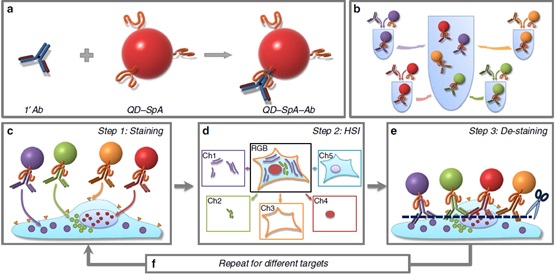
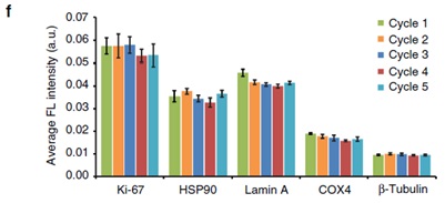
Title: Nature communications : Multi-round multicolor in situ imaging based on quantum dots Abstract: Based on the quantum dot-Protein A-antibody conjugate, multiple rounds of multi-color co-staining of the same sample were performed, and the fluorescence spectrometer was used to analyze the potential of 50-100 target molecules in a single cell. The Gao Xiaohu group of the University of Washington used the conjugate of Protein A and Quantum dots (QDs) to capture the antibody Fc fragment by Protein A to obtain the QD-Protein A-Ab fluorescent probe (Fig. 1). The quantum dot fluorescent probe is simple in preparation, low in cost, suitable for pipeline analysis, and easy to elute, does not interfere with the next round of staining, and maintains the integrity of the target antigen and specimen morphology in each round of staining. For each round of multi-color co-staining, it is necessary to use fluorescence spectrometer for spectral separation and quantitative analysis to obtain detailed molecular quantitative information of single cells, which is important for system biology, gene expression research, signal transduction pathway analysis and molecular diagnosis. significance. In each round of multicolor co-staining, N target molecules can be analyzed, and the size of N depends on the spectral resolution of the different fluorescent probes. If there is a corresponding quantum dot for antibody labeling at every 20nm emission wavelength, and the fluorescence spectrometer has ten channels for spectral separation and quantitative analysis, then ten colors of co-staining can be performed simultaneously in each round, if another nine rounds are passed Decolorization-re-staining, theoretically, enables analysis of a total of 100 (10X10) target molecules on the same fixed sample. The above research group carried out in-situ imaging analysis of 5 colors of co-staining and repeated 5 rounds in each round. The results showed that all five target molecules showed consistent dyeing patterns and average staining intensity in five rounds of dyeing, and each After the round of staining, no fluorescence signal remained, and the specimen maintained a good cell morphology after five rounds of staining (Fig. 2). Figure 1 Multi-round multicolor in situ imaging pattern of QD-Protein A-Ab fluorescent probe (a) Prior to staining, the QD-Protein A conjugate captures the antibody (Ab) in solution to form a functionalized QD-Protein A-Ab fluorescent probe; (b) pools the different probes into a cocktail mixture; Hybrid probes are incubated with fixed cells for parallel multicolor co-staining; (d) Image and spectral data are acquired using a fluorescence spectrometer to form expression profiles of each antigen; (e) Brief rinsing with regeneration buffer to complete the specimen Decolorization; (f) Multicolor co-staining of the next round of other targets. Figure 2 25 target imaging analysis of QD-Protein A-Ab fluorescent probe (ae) Five rounds of target imaging: QD-Protein A-Ab with an emission wavelength of 525/545/565/585/605 nm, respectively labeled Ki-67/HSP90/Lamin A/Cox-4/b-tubulin); (f) Five rounds of staining have consistent average fluorescence intensity. Source of the document: Zrazhevskiy P, Gao X. Quantum dot imaging platform for single-cell molecular profiling. Nat Commun. 2013, 4:1619. Isomalto-Oligosaccharide 900 (tapioca) Syrup Isomalto Oligosaccharide Prebiotic Fiber,Prebiotics Supplements,Short-chain Carbohydrates Shandong Bailong Chuangyuan Bio-tech Co.,Ltd. Qingdao Branch , https://www.qdblcycn.com